Cervical Spine
The spine or spinal column (commonly called the backbone) is that part of the skeleton that runs from the skull above to the pelvis below. It is divided into five regions from top to bottom: the cervical spine in the neck, the thoracic spine corresponding to where the ribs arise, the lumbar spine in the low back, the sacrum at the base of the column and in between the two halves of the pelvis, and the coccyx hanging off the end of the sacrum (essentially all that remains in humans of what would be a tail in other species).
The spinal column is made up of a number of individual bones called vertebra which stack up one on top of the other and are joined together by ligaments, muscles, and a specialised cartilage called the intervertebral disc. The disc functions to tether one vertebra to the next but also as a shock absorber for the motion that occurs between neighbouring bones. Each vertebra has a large hole in its middle which, when joined up with that of the neighbouring vertebrae, together form a tube up and down the spinal column which is called the spinal canal.
There are usually seven vertebrae in the cervical spine, twelve in the thoracic, and five in the lumbar region. Those in the sacrum have fused together so that the original five here form one complete bone. The coccyx is rudimentary in humans and can consist of three to five bones. There can be some variations of “normal” here with so-called “segmentation anomalies” (for example, the top sacral vertebra may not be joined to the rest giving the appearance of six lumbar vertebrae). The presence of a segmentation anomaly is of no functional concern to an individual patient other than that the treating surgeon needs to recognise it when making certain diagnoses and planning treatment.
Looking from the front the normal human spine is usually very straight, although looking from the side there are three broad curves with the cervical spine in the neck being curved gently backwards (so called “lordosis”), the thoracic spine curved forwards (“kyphosis”), and the lumbar spine again curved backwards into lordosis. These curves balance each other out so that in the young adult spine the skull is position vertically over the pelvis. With aging we commonly see the head seeming to creep forward in front of the pelvis often due to “loss of lordosis” in the cervical or lumbar regions or “exaggerated kyphosis” in the thoracic spine. Although this can be a normal part of aging certain diseases, injuries, or degenerative process may lead to abnormal curves resulting in “loss of spinal balance” that may require treatment.
The pattern of vertebrae and intervertebral discs repeats throughout the vertebral column with each vertebra being named by region and number. The first vertebra in the lumbar region therefore is called “L1”, the fifth in the thoracic region T5, the third in the cervical region C3, and so on. Each intervertebral disc is named by reference to its two vertebral neighbours, so that the disc in between L4 and L5 is called the L4/5 disc, that in between L5 and S1 is called the L5/S1 disc (or less commonly the “lumbosacral disc”), that between C6 and C7 the C6/7 disc, etc. Since the sacrum is one complete bone there are no sacral discs, and the top two vertebrae in the neck (C1 and C2) are also very specialised to allow the high degree of movement between head and neck so that there are no discs here either. The highest disc level therefore is the C2/3 disc connecting C2 to C3, and the lowest is the lumbosacral disc at L5/S1.
Between each pair of vertebrae a spinal nerve (also called the nerve root) will exit from the spinal column. These are named from top to bottom so that the C1 nerve is the first to leave below the skull and above C1, then C2 coming out above C2, the C3 nerve above the C3 vertebra, etc. Things change a little at the junction between the cervical and thoracic spine at the base of the neck with the C7 nerve exiting above C7 as usual but then the C8 nerve coming out below the C7 vertebra (there are only seven bones but eight nerves in the cervical spinal column). The pattern is then consistent down the rest of the spine with each nerve leaving below its equally named vertebra so that the T1 nerve comes out below T1, T2 below T2, and so on all the way down to and through the sacrum. A spinal segment corresponding to one vertebra, its neighbour, their intervening intervertebral disc and the exiting spinal nerve is known as a spinal “level”.
Each spinal nerve will eventually look after a specific region of the body. That area where it supplies sensation is referred to as a “dermatome”, whereas that region where it controls muscle movement or motor control is called a “myotome”. Some of the spinal nerves (although not all) will also supply the so-called deep tendon reflexes such as the knee jerk reflex that a doctor may try to elicit with a tendon hammer. The importance here is that with careful assessment of sensory, motor, and reflex symptoms and signs the neurosurgeon can often identify which spinal level may be the site of a problem.
Common conditions throughout the spine
Herniated Intervertebral Disc
The intervertebral discs are the specialised cartilages that sit between two neighbouring vertebral bones. They function both as ligaments to help regulate the movement of one bone on the other, and also as shock absorbers that help to spread the load of the body weight in the upright human posture. Like all cartilages in the body they are prone to wear and tear degenerative changes (part of spondylosis) and also to true injuries.
A disc is said to herniate if the tough outer lining of the disc develops a tear that allows some of the softer inner component to “prolapse” or “slip” out. Sometimes this may just be a general bulge or on other occasions may be a free or so-called “sequestered” fragment. Often, this may cause little to no symptoms, but should the disc compress the underlying nerve or spinal cord it causes significant pain and loss of function.
Over time the body can heal disc herniations spontaneously, although not always. Initial treatments therefore are usually non-surgical in order to try and foster this healing process such as physical therapies, medications, injections, or just managing and allowing time to pass. If the condition is not improving or if there is evidence of worsening pain or loss of function, then surgery may be required to remove the herniated portion of the disc in order to decompress the underlying nerve tissue.
Spondylosis
This is the name given to the age related wear and tear changes that occur in the spine. It is not truly a disease since it will happen to everyone who lives into adult life, similar to going grey of the hair and developing ageing of the skin. It involves a combination drying out of cartilages, adjacent bony spur formation, discs becoming collapsed and bulging, ligaments losing their elasticity and seeming to thicken, sometimes degenerative cyst formation, and joints becoming unstable and incompetent. It is very similar to degenerative arthritis elsewhere in the body such as osteoarthritis. From person to person it can occur at different rates and at different ages related to genetic factors, heredity, previous injuries, and other conditions that may load the spine such as carrying around too much body weight, repetitive lifting and twisting movements over the years (e.g. so-called “builders’ or nurses’ back”), etc.
Where it differs from arthritis elsewhere however is that the combination of these changes can result in extra tissue being laid down which can then take up the space that is needed for either the spinal cord or the spinal nerves. This narrowing of the spinal space is what is referred to as “spinal stenosis”.
Since spondylosis itself is not a disease there is no definite treatment, and generally it is recommended to look to modify risk factors to try and reduce the rate that it progresses (e.g. weight reduction, identification and correction of errors of posture, avoiding wherever possible repetitive lifting and twisting manoeuvres, and core strengthening exercises).
However, if spondylosis occurs to too great a degree and the extent of the stenosis is enough to jam the nerves or the spinal cord then it can lead to the conditions of spondylotic radiculopathy (if one nerve is jammed), neurogenic claudication (if many nerves are jammed particularly in the lumbar spine), and spondylotic myelopathy (if the spinal cord itself is jammed particularly in the neck).
The Spinal Canal and Spinal Canal Stenosis
The spinal canal is the name given to the roughly tubular space that runs up and down the centre of the spinal column and through which run the spinal cord, the spinal nerves and their coverings (including the cerebrospinal fluid or CSF). It is created by each individual vertebra bone having a large hole behind its weight bearing part (or vertebral body). With each vertebra lining up one on top of the other (separated by the intervertebral discs) this large central hole connects up to its neighbour and so forms a canal along the length of the spine.
The width and shape of the spinal canal varies from person to person in a normal way similar to other physical features such as body shape and height. If the spinal canal is on the narrow side of normal we sometimes refer to this as the person having a “congenitally narrow spinal canal”. Although this is not a problem in itself it can set the scene for potential nerve or spinal cord compression in the future as the canal develops further narrowing with age due to the process of spondylosis (described above). The combination of acquired or so-called spondylotic spinal canal stenosis on the background of a congenitally narrow spinal canal may well be something that brings a patient to the attention of a neurosurgeon. Although everyone will develop some degree of spondylosis and canal narrowing over time those of us with a wide canal to begin with may never get enough narrowing over the course of their life to develop nerve or spinal cord compression.
The spinal cord occupies roughly the top two thirds of the spinal canal, finishing at roughly the L1 level in most people. In the low back therefore there is no longer any spinal cord and the canal is occupied by all of the spinal nerves running down to exit at each level on their way to various parts of the leg and pelvis. These nerves together are sometimes called the “cauda equina” since they reminded the early anatomists of the hairs on a horse’s tail.
In the lumbar spine the canal become more triangular in shape, and in some people may even adopt a little clover leaf or “trefoil” shape. The side part or the arm of the triangle is referred to as the “lateral recess” of the spinal canal.
At the junction between each vertebra (a so-called “level”) a spinal nerve will travel out of the spinal canal through its own hole into the body on its way to look after a specific area (generally part of the arm for the cervical nerves in the neck, the trunk for the thoracic nerves in the middle back, and the leg and pelvis for the lumbar and sacral nerves in the low back and tail bone). These individual holes are called the “nerve root exit foraminae”.
All of these areas are prone to develop the process of spondylosis which can lead to extra thickened tissue being deposited over time. This may occur to such an extent that these spaces can become narrow leading to the description of spondylotic spinal canal stenosis. You may hear terms like “central spinal canal stenosis”, “lateral recess stenosis”, or “foraminal stenosis” referring to narrowing of these specific sites in the spinal canal which can have implication as to which if any of the nerves or the spinal cord may become compressed. The lateral recess is particular prone to stenosis since its roof is formed by large joints called facet joints which allow one vertebra to move on top of another. These joints are common sites of the wear and tear type changes that can result in the bony spurs, degenerative cysts, or thickened ligaments can make up the process of spondylosis. Since a spinal nerve often runs down through the lateral recess on its way into its own exit foramen this is a common site where nerve entrapment may occur.
Cervical Spine Conditions
The Cervical spine is that part of the vertebral column located in the neck. It is a common location to develop problems due to the large amount of motion that normally occurs in this region of the body. It is also prone to wear and tear or “degenerative” changes as well as true injuries and is an area that often suffers from long term postural errors in our modern desk and screen oriented world.
Like the rest of the vertebral column the cervical spine consists of individual bones (or vertebra) connected to each other via ligaments, muscles and specialised cartilages called intervertebral discs. There are 7 individual bones in the neck with the top 2 (C1 and C2) being very specialised and not following the “normal” pattern. All these bones move one on top of the other in a very regulated way. Age, disease, or injury can in some cases effect this pattern and may result in abnormal motion which we call “instability”.
It is a delicate and important part of the human body due to the presence of the spinal cord and the cervical spinal nerves. The spinal cord is that part of our nervous system which allows us to move and feel with our bodies. It runs down through the central canal of the spinal column that is made by each vertebra joining up with the next. Diseases or damage here can be very serious leading to significant disability such as paralysis (quadriplegia of involving all four limbs, paraplegia if involving just the legs).
The spinal nerves connect the spinal cord with the body. At each space between one vertebra and the next (which we call a “level”) one spinal nerve will leave the spine on each side and end up travelling to its own specific part of the body. The hole through which the nerve leaves the spine is called the “exit foramen”, and this part of the nerve is often referred to as the “nerve root”. Those in the neck are mostly involved with the function of various parts of the arm and hand.
In certain situations, the spine may develop narrowing of the spaces for the nerves or spinal cord. In medicine we call this narrowing “stenosis”. It is often caused by the wear and tear changes in discs, ligaments, and bones that can develop over time in an aging process similar to arthritis that we call “spondylosis”. If this stenosis occurs to too great a degree it can take up the space needed by the spinal cord or the spinal nerves leading to these structures becoming damaged by being crushed or “compressed”.
Many common problems in the cervical spine can be successfully treated without surgery, for example with physiotherapy or other gentle manipulative treatments, exercise, medications, or injections. However, these treatments may not always be effective and may be too risky to persist with or try, for example if they are not likely to reverse the underlying problem or that problem is progressing despite these treatments.
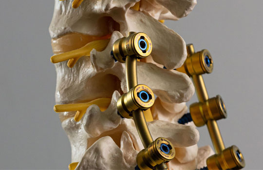
Surgery in the cervical spine may be needed for many reasons. Most commonly it is designed to “decompress” these vital structures in order to prevent further damage from occurring and to foster the recovery of any existing damage. Other reasons include the need to re-stabilise the spine if instability has developed, correct deformity, treat fractures, remove tumours, treat infections, etc.
Common conditions that may require surgery include cervical radiculopathy and cervical myelopathy.
Unfortunately, there are also some common problems for which surgery may not be that beneficial, e.g. neck pain or whiplash. It takes a careful and thorough assessment to know when surgery may help, and a degree of wisdom to know when it may just be meddlesome. A good surgeon not only knows how to perform surgery but also knows when it should be offered or not. Sometimes the best way a skilled surgeon can offer a patient is to explain why surgery may not be a good option of treatment in their particular case.
Common conditions requiring surgery:
Cervical Myelopathy
This occurs when the spinal cord in the neck becomes squashed for example by a large herniated cervical disc or by spondylosis. Left untreated the condition often progresses with damage occurring in the spinal cord that may not fully recover even after the compression is relieved. It is important therefore to detect and treat this condition early before significant disability develops.
Common symptoms include numb, clumsy hands, loss of dexterity (difficulty typing, texting, manipulating small objects such as doing up buttons, worsening handwriting, etc), stiff, slow and unsteady walking particularly on uneven ground, slopes, slippery surfaces etc. Early symptoms may be mistaken for other co-existing conditions such as hand or knee arthritis. Symptoms may evolve slowly over time (months or years) but can develop more rapidly often in a step wise pattern with an apparent loss of function then seeming to stabilise over time before another loss occurs. As time passes the size of these steps often increases. This is a serious condition that left untreated can result in the need for wheel-chairs and significant loss of independence with basic functions such as eating, dressing, toileting, etc.
There is no one single treatment for this condition with each case needing a careful assessment to determine exactly what needs to be done. In general however the principals of treatment include decompressing the spinal cord, restabilising the spine and correcting deformity (if needed).
Cervical Radiculopathy
This refers to symptoms due to a problem with one of the nerve roots in the neck, most commonly the nerve being squashed by a herniated disc or due to spurs that develop with spondylotic foraminal stenosis. Generally, the symptoms are experienced not just in the neck but more so in that part of the body that the specific nerve involved looks after. For nerves in the neck this mostly means in one part or other of the arm. Symptoms can include a combination of one or more of pain, pins and needle, numbness, or weakness and vary according to exactly which nerve is involved.
Initial treatments are usually non-surgical including time, medications, physical therapies, traction, injections, etc. However, surgery may be needed if these treatments are not working over a reasonable period of time (with “reasonable” depending on how bad and exactly what the symptoms are), if the symptoms are getting worse despite these treatments, or of very frequent flare ups occur.
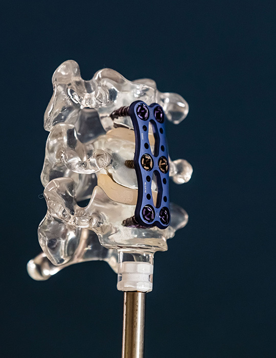
There can be many types of surgery offered for this condition including posterior cervical foraminotomy, microdiscectomy, anterior cervical discectomy and fusion, or artificial disc replacement. A careful and thorough assessment is needed to work when surgery is needed and what type would be the best option in the individual case. Dr Brennan works according to the general principle of looking for the least invasive option to treat the individual condition at hand.

