Minimally Invasive Surgery
General Anatomy and Function
Most modern operations are less invasive now than they were in the past. Minimising tissue disruption and limiting the need for reconstruction leads to faster recoveries and less post-operative discomfort. In many situations a minimally invasive approach has been made possible due to the development of new devices such as retractor systems, operating microscopes, and intraoperative imaging (Xray, CT, and MRI). More recently, spine navigation (in which there is real time feedback correlating the surgical anatomy with the CT or MRI scan) and robotics has further enabled an increased precision.
- Retractors have been developed which can very efficiently maintain the exposure of the operative corridor through relatively small incisions. This can make “access” to the spine possible with much less disruption of muscle and tendon then previously.
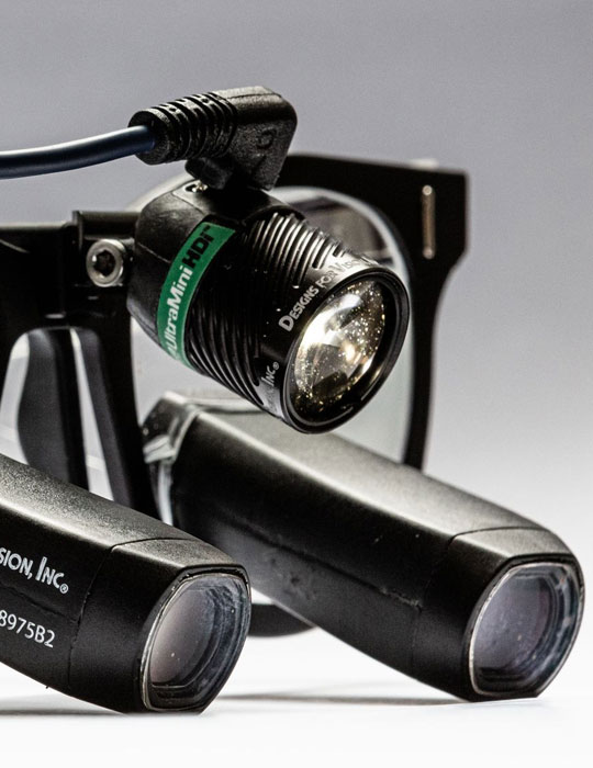
- The operating microscope is key to minimal invasive surgery as it allows brilliant illumination and a high degree of magnification of the operative field. Unlike some other surgical specialties where a microscope may be an unfamiliar piece of equipment for a neurosurgeon who has been trained in micro-neurosurgery in the brain the use of the microscope in the spine is a natural extension of skills to improve the fidelity of minimally invasive spine treatment.
- Imaging at the time of surgery is critical when making minimally invasive approaches in order to know precisely where and at what level in the spine the exposure and subsequent surgery is being made. Traditionally this has been done with Xrays (real time fluoroscopy or image intensification). This equipment provides views of the target zone at a number of different angles, but each view is limited to two dimensions due to the technology involved. For most procedures on table fluoroscopy remains an important tool at the surgeon’s disposal.
More modern advances in imaging however include on table CT scan or CT-like imaging. These have the advantage of providing cross-sectional pictures in addition to the standard side-on or front-on views of 2-D Xrays. This can be useful in imaging the spine in cases where standard fluoroscopy may be difficult to interpret such as if there is significant deformity, previous surgery, or other patient conditions such as very weak bones from osteoporosis (which may be difficult to show up on fluoroscopy) and if there is more soft tissue in the field that can absorb and scatter the xray beam (such as can in cases of increase obesity). It can also be particularly useful in checking that any implants that may have been required are placed precisely where they need to be. In the rare situation where the position of an implant can be improved it allows this to be done then and there since the location of the implant is determined while the operation is still proceeding rather than on the post-operative scan the next day. They can take more time to use and, in some situations, may expose the patient and theatre staff to a higher dose of Xrays so their use needs to be planned appropriately.
Intraoperative MRI scan can be very useful for brain surgery, but currently has a limited role in the spine.
- Navigation refers to the ability to have a (usually) visual feedback as to precise location when performing surgery so as to increase precision, particularly in situations where direct visualisation of the target is more difficult (such as very deep locations, locations that are not directly exposed, and in minimally invasive procedures where views may be limited). This feedback is usually displayed on a monitor screen during the operation and with reference to either the pre-operative imaging, the on table CT or CT-like scan, or some combination of the above. It is very similar to a GPS navigation device in the car or smart phone in that although the driver or paedestrian may know where they are the navigation can give an overview of where they need to be and how to get there.
Neuronavigation is now standard in many brain surgery procedures and is becoming increasingly useful in spine surgery. Neurosurgeons therefore are very familiar with its use across these regions of the body as well as with how to best augment their existing surgical skill and technique with this technology. It can be particularly helpful in some situations when placing spinal implants during a spinal fusion procedure, particularly in the lumbar spine.
Combining navigation with on-table imaging provides a way to constantly update the “GPS map” which can alter as things may move around during the course of an operation.
- Robotics is currently in the early generation of development for spinal surgery. In certain situations a robot can increase in the precision of placing certain instrumentation such as pedicle screws. There is promise for future applications as this technology grows.
In some situations however a small opening may not always be the safest. Neurosurgery differs from most other surgeries due to the delicate and unforgiving nature of the brain, spinal cord and nerves, and therefore the safest operation is the one which will allow the greatest “control” of these tissues to maximise chances of success and minimise risks. It must always be remembered that a skin incision will heal no matter its length, but injury or damage to the nerve tissue of the brain and spine may not.

